-
Fluorescent imaging training
Part 1: basics
Welcome to our training series on fluorescent imaging. We’ll show you imaging basics and essential steps before moving on to optimization, troubleshooting, and more advanced techniques.In Part 1 of our series on fluorescent imaging, we introduce you to some basic but essential tools and concepts in order for you to get the most out of your fluorescent imaging, especially when it comes to imaging cells.
Part 1 overview
1.1 Using a hemocytometer
1.2 Aseptic technique and clean cultures
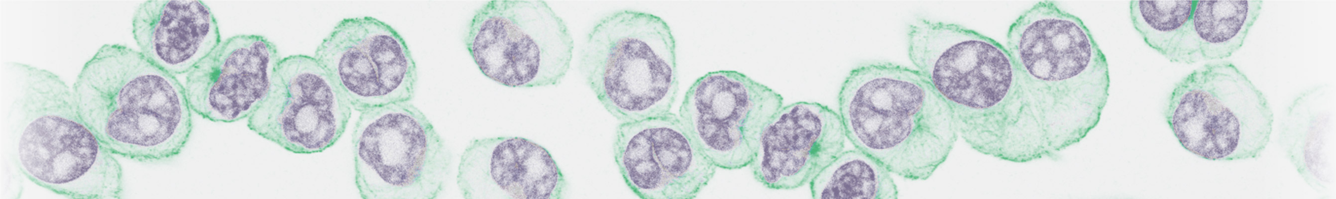
1.3 The confocal microscope
1.1 Using a hemocytometerCell counting is an important step if you're using fluorescent imaging to assess the extent of a change in your cell populations. One of the best ways to quantify your cells is with a hemocytometer. Here's a great video that covers how to count suspension and adherent cells. You can get a downloadable written protocol of this here.
1.2 Aseptic technique and clean culturesProper aseptic technique is essential to avoid contaminating your cell cultures and reagents. In this video, we outline our protocol to sterilize work areas and use appropriate sterile handling techniques, personal protective equipment, and, of course, good hygiene. If you'd prefer a written protocol, you can find it here.
1.3 The confocal microscopeA confocal microscope is an incredibly powerful tool that we commonly use for fluorescent imaging of both tissues and cells. There can be quite a lot of complexity to deal with when you first use this piece of equipment, so in the webinar, we walk you through the major steps. Here you'll learn about sample preparation at the very beginning, through to data analysis, common pitfalls and how to overcome them. Finally, we give you a rundown of how to optimize your setup and a few of the advanced applications that make use of confocal microscopy.
SummaryYou should now have a better understanding of how to
– Get a good cell count using a hemocytometer
– Work with good aseptic technique
– Use the confocal microscope
In Part 2, you'll learn about choosing the right antibodies for fluorescent imaging, as well as the pros and cons of direct versus indirect staining.